Categories
- Cardiology 01
- COVID-19 68
- COVID-19 Treatment 68
- Gastroenterology 02
- Health Care 02
- Hospital 01
- lymphedema 72
- Neurology 01
- Orthopedics 01
- Peripheral Arterial Disease 02
- Prescott vein clinic 108
- Pulmonology 03
- Radiology 03
- sclerotherapy 91
- skin ulcers 90
- Uncategorized 51
- Varicose Veins 107
- vascular surgeon 149
- vascular surgeon in Phoenix, AZ 147
- Vein Clinic 146
- vein clinic in arizona 148
- vein disease 146
- Venous Insufficiency 96
- Venous Ulcers 93
Recent Posts
Deep Vein Thrombosis (DVT): Symptoms, Causes, and Prevention
When Should You See a Vein Specialist? Key Symptoms to Watch For
Case Study: Lower-Limb Venous Duplex Ultrasound Examination
Evaluating Leg Swelling and Venous Circulation
Patient Overview
A 62-year-old female presented with persistent swelling, heaviness, and aching in her left leg. The symptoms had gradually increased over several months and were more noticeable after prolonged periods of standing.
The patient also reported visible veins around the lower leg but had no known history of deep vein thrombosis (DVT).
The Clinical Concern
Because persistent leg swelling can have several possible causes, including venous insufficiency and DVT, a Lower-Limb Venous Duplex Ultrasound Examination was recommended.
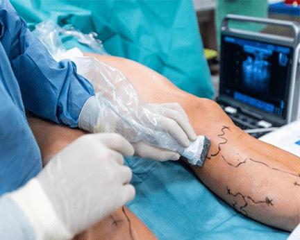
The examination was performed to assess blood flow through the deep and superficial veins, check for venous obstruction or blood clots, and identify any abnormal backward flow of blood known as venous reflux.
Ultrasound Examination
The venous duplex ultrasound provided real-time images of the veins in the affected leg. The examination evaluated:
- Deep and superficial venous systems
- Venous blood flow
- Vein compressibility
- Evidence of obstruction or thrombus
- Venous reflux
- The great saphenous vein and other clinically relevant vessels
Findings
The ultrasound showed venous reflux involving the superficial venous system, consistent with venous insufficiency.
The deep veins remained patent and compressible, with no evidence of acute deep vein thrombosis.
The findings helped explain the patient’s ongoing leg swelling, heaviness, and visible varicose veins.
Diagnosis
Chronic Venous Insufficiency with Superficial Venous Reflux
No evidence of acute Deep Vein Thrombosis (DVT).
Treatment Plan
Based on the examination findings and clinical assessment, the patient was advised on conservative measures and further treatment options.
The initial recommendations included:
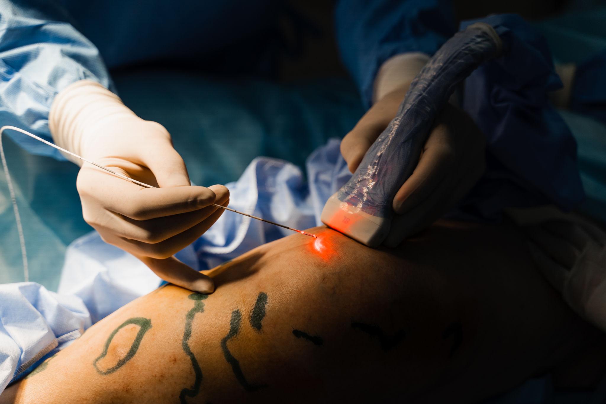
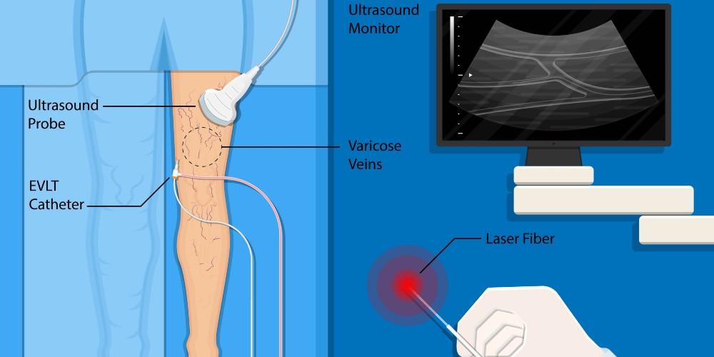
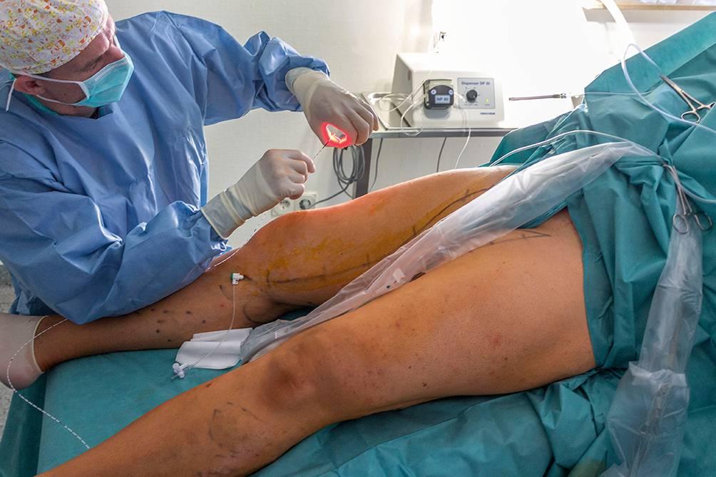
For persistent symptomatic reflux, an appropriate minimally invasive venous treatment may be considered following evaluation by a qualified vascular specialist.
Outcome
At follow-up, the patient reported improvement in leg heaviness and discomfort after following the recommended management plan.
The duplex ultrasound findings also provided an important baseline for monitoring the patient’s venous circulation and guiding future treatment if symptoms persisted.
Why Venous Duplex Ultrasound Matters
A lower-limb venous duplex ultrasound is a non-invasive examination that can provide valuable information about blood flow and the condition of the veins.
For patients experiencing unexplained leg swelling, heaviness, aching, varicose veins, or other symptoms associated with venous disease, appropriate vascular imaging can help identify the underlying problem and guide the next step in care.
Important: This case study is for educational purposes only. Individual symptoms, ultrasound findings, diagnosis, and treatment should always be evaluated by a qualified healthcare professional.
General Ask
What is a Lower-Limb Venous Duplex Ultrasound?
Why might I need a Venous Duplex Ultrasound?
Can a Venous Duplex Ultrasound detect a blood clot?
Is a Lower-Limb Venous Duplex Ultrasound painful?
How long does the examination take?
Conclusion
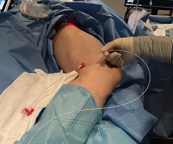
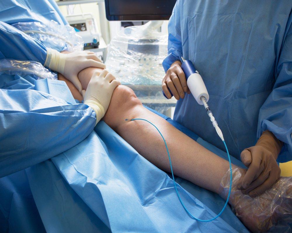
Endovenous Laser Treatment (EVLT) is an established minimally invasive approach for appropriately selected patients with symptomatic varicose veins and superficial venous reflux. Careful duplex ultrasound assessment, ultrasound-guided treatment and appropriate patient selection are essential components of successful management.
In this illustrative case, EVLT provided a minimally invasive method of treating the underlying incompetent vein while avoiding conventional surgical vein stripping. Current NICE guidance places endothermal ablation among the preferred interventional treatments for suitable patients with confirmed truncal reflux.
Clinical disclaimer: This case study is intended for educational/healthcare communication purposes and is not a substitute for individualized medical assessment. Actual patient information, procedural parameters and outcomes should be documented from the clinical record rather than inferred from the photograph.